ABSTRACT
Background
The study aimed to evaluate the potential of Momordica cymbalaria Fenzl (MC) in healing burn wounds. Partial-thickness scald wounds were induced on SD rats by pouring hot molten wax at 800°C on the dorsal area of the rat. Rats were placed into five groups (n=6). 1st group served as the wound control (Untreated) group. The rest of the 4 groups received SSC (1% w/w), 5% extract of MC, Scaffold, and 5%, Saponin of MC topically respectively twice daily for 14 days. Wound contraction and period of epithelization were checked on days 1,9 and 14. On the 9th day, the granulation tissue was isolated and estimated for hydroxyproline content and antioxidant enzymes. A part of the granulation tissue was processed for histopathology studies.
Results
5% extract and Saponin of Momordica cymbalaria Fenzl promoted wound contraction and decreased the period of epithelization when compared with control but lesser than the SSC-treated group. It also enhanced collagen synthesis and wound stability, as demonstrated by an elevation in hydroxyproline content. The histological examinations also affirmed the healing potential of the drug, which showed a full form of epidermis layer, less inflammatory cells, more no. of collagen fibers, and blood vessels lesser than the SSC-treated group and scaffold. Endogenous enzymatic and non-enzymatic antioxidant levels increased significantly in the Momordica cymbalaria Fenzl -treated groups, while lipid peroxide levels decreased.
Conclusion
By 5% extract, Momordica cymbalaria saponin, and scaffold have great healing capacity in burn wounds and have a beneficial influence on the various stages of wound restoration.
INTRODUCTION
Burns can be considered a global health problem, which could result in trauma and devitalization of tissues or organs leading to morbidity and mortality.1 As per the World Health Organization (WHO) burns are estimated to cause 180,000 deaths annually. Worldwide, around 11 million burn victims in 2004, needed medical treatment.2 Every year, more than 100,000 people in India suffer mild to severe burns.3 Wound healing is a synchronized mechanism that integrates multiple phases like homeostasis, the Inflammatory phase,4 the proliferative phase,5 and the remodeling phase.6
Ayurveda and Siddha, the most ancient traditional systems of medicine, are explored once again. Future medical systems are also being connected, as natural remedies are taken from mother nature and put together with cutting-edge technology to create a readily available treatment. Plant-based medications despite being around for 5000 years have not been exploited to their true potential until recent years, in developing countries approximately 70–90% population is shifting towards the ancient remedies constituting plant extracts to a major proportion.7
One-third of medications out of 1-3% representing medication for wounds on the skin in the western pharmacopeia are plant-derived.8 Several studies have proved that phytochemicals from plants act through multiple mechanisms leading to the healing and formation of new tissues in the affected area.9
Saponins are glycosidic secondary metabolites present in plant species but also in certain animal sources such as marine crustaceans.10 The role of triterpenoid saponins in wound healing has been thoroughly investigated through various studies. Studies by Kimura et al. with Asiaticoside11 and Madecosside,12 a triterpenoid saponin obtained from Centella asiatica (Umbelliferae), demonstrated its anti-inflammatory effect in rat scald models. The antioxidant and metalloprotease activity of Bacoside A extracted from Bacopa monnieri showed potential in tissue remodeling after burn injury.13 These results concluded the significance of triterpenoid saponins in wound healing caused by burn injury via inhibiting apoptosis14 and ROS-scavenging activity.13
Many phytochemicals that have the potential for wound-healing activity have been discovered and employed in the modern market. Momordica cymbalaria (synonyms; Momordica tuberosa Roxb. Cogn., or Luffa tuberose Roxb.) is a member of the Cucurbitaceae family. Different extract of the crude drug is said to possess anti-diabetic, hypoglycemic,15 anti-diarrheal activity,16 anti-cancer17 and anti-microbial activity,18 anti-ovulatory and abortifacient potential,19 hepatoprotective and antioxidant activity.20
Since the tissue engineering era, scaffold, 3D structures that resemble the extracellular matrix of living tissue have been assessed for different functions including wound healing and regeneration aspects.21 The properties to be considered during the employment of the scaffold in wound healing is a suitable mechanical and physical characteristic and a strong physiological foundation that promotes cell adhesion, proliferation, and/ or differentiation, They must have a degradation profile that corresponds to the time needed for wound healing and be both biocompatible and biodegradable. High porosity, a high surface area to volume ratio, an interconnected architecture, and the flexibility to conform to the contour of the wound.22
The current study was conducted to demonstrate the methanolic effect of MC and the saponins isolated from methanolic extract on burn wound healing and also its antioxidant properties. The study also compares with the scaffold23 (commercial bio-engineered product).
MATERAILS AND METHODS
Chemicals and reagents
Ehrlich’s reagent, Poviz-MA, Xylazine hydrochloride, and Ketamine hydrochloride intraperitoneally, Ehrlich’s reagent, soft white paraffin.
Preparation of extract and isolation of saponins of MC
Momordica cymbalaria roots were collected and minced, before being dried in the shade for seven days at room temperature. The roots obtained after drying were pulverized and sieved through Coarse10/40 sieve. The extract was prepared by boiling 70% Me OH with dried powder of Momordica cymbalaria (1.0 kg) in reflux condition.24 After filtration, the solution was condensed under a vacuum. Momordica cymbalaria methanolic extract was dissolved in hot distilled water, then partitioned (1:1) between the water layer and n-butanol saturated with water. Separation and evaporation of the organic layer (n-butanol layer) yielded the residue, which was further dissolved in methanol and poured in diethyl ether (Et2O) (1:1) to obtain a flocculent precipitate. The crude saponin fraction was produced after this precipitate was separated using filter paper, washed with excess ethanol, and dried.25 The saponin mixture was then dissolved in distilled water and utilized for investigation.1,27
Formulation of ointment
Using soft white paraffin, ointments of 5% w/w were created.28
Animals
The Institutional Animal Ethics Committee granted ethical clearance for the study protocol. Sprague-Dawley (SD) rats of either sex (150-200 g) procured from NIMHANS, Bangalore, were used for the study. According to laboratory practices, the animals were acclimatized for 10 days. The rats were placed in polypropylene cages and kept at a relative humidity of 65 ± 10%, 12 hr of light/dark cycle, and at 27 ± 20°C. They were provided with water ad libitum and a rodent pellet diet (Gold Mohur Lipton India Ltd.,). Dermal toxicity performed according to (OECD guidelines).
Burn wound model (hot wax model)
Healthy adult SD male rats (150-200 g) were subjected to burn wounds of partial thickness. Animals were divided into a group of 5, 6 animals per group with full access to pallet diet and water.
Each Rat was administered 50 mg/kg of Ketamine Injection (IM) to induce anesthesia, and electric clippers were used to trim the fur on the back. A metal cylinder, (300 mm2 areas of circular openings with a capacity to hold 4.0 g of wax) were carefully placed on the trimmed area, and burn wounds were produced by pouring hot molten wax at 80°C. The metal cylinder with the wax attached to the skin was removed when the wax solidified (8 min), leaving 300 mm2 of well-defined circular incisions. Before being transferred to holding areas, animals subjected to the procedure were housed in separate cages to recuperate fully from the anesthetic.27
Drugs Treatment and Groups Division
Group 1: White Soft Paraffin 2 times a day (Wound Control).
Group 2: Silver Sulfadiazine Cream (SSC) (1% w/w) mg/kg.
Group 3: 5% Methanolic Extract of MC 5 mg/kg.
Group 4: 5% Saponin of MC 5 mg/kg.
Group 5: Scaffold mg/kg.
Treatment for all the groups was made twice a day for 14 days. Daily observation of animals was conducted and the extent of the healing was determined using the physical parameters, the epithelialization period, and the contraction of the wound.
Wound contraction
Planimetric assessment of wound progression was performed, omitting the day of injury induction. Measurement of wound size was performed daily. In the following equation, the assessed surface area was used to compute the percentage of wound contraction, considering the original size of the wound, 300 mm2, as 100%.29
Wound contraction = (Initial size of the wound -wound size of the specific day)/(Initial wound size) X100
Epithelialization Period
It was evaluated by keeping track of how long it took for the eschar to peel away from the burn/Scald site surface without producing a raw wound.30
Biochemical Parameters like Hydroxyproline Content1,32 and Total Protein Estimation (MM, 1976) were performed by using the granulation Tissue Excised on the 8th-day post-wounding. Lowry’s method was followed to determine the protein content in the sample.
Antioxidant activity
Lipid Peroxidation (LP)33
The level of Malondialdehyde (MDA) was used as an assessment parameter to determine the degree of lipid peroxidation in tissues, as per Wilbur et al.
Superoxide Dismutase (SOD)34
SOD measurement was done according to the method of Kono et al.
Catalase (CAT)35
Catalase activity is estimated based on the breakdown of H2O2 at 240 nmin in an assay combination including phosphate buffer and is expressed in units as µM of H2O2 consumed/min/mg of protein.
Glutathione-s-transferase (GST)36
The assay was conducted using Habig et al. procedures, with a few minor modifications made in accordance with Anosike et al.
Reduced glutathione (GSH)37
Ellman’s method was used for the GSH assay.
Histological processing and microscopic analysis
The animals were euthanized using anesthetic diethyl ether and their skin was dissected out. The wound biopsies were fixed in 10% neutral formalin for at least a week before they were sectioned with a cryotome into 5µm sections. The processed sections were protected by coverslips and were studied with a light microscope for histopathology analysis.
Statistical analysis
Results were expressed as mean ± standard error means by using one-way ANOVA and Dunnett’s comparison between wound control (Untreated) v/s all groups. The data were considered significant at p<0.05.
RESULTS
Effect on Wound contraction
Application of Silver Sulfadiazine Cream (1% w/w) to the hot wax burn wound produced significant contraction of the wound (p<0.001) on the 9th day when compared to untreated hot wax burn wound rats. Whereas when Group 3 was compared to the untreated control resulted in significant (p<0.01) wound contraction on the 9th day.

Figure 1:
(A) Histogram representing Hydroxyproline content (B) Histogram representing Total protein

Figure 2:
Histogram representing antioxidant activity (A) Catalase, (B) Superoxide dismutase, (C) Glutathione-s-transferase, (D) Reduced Glutathione, (E) Lipid peroxidase

Figure 3:
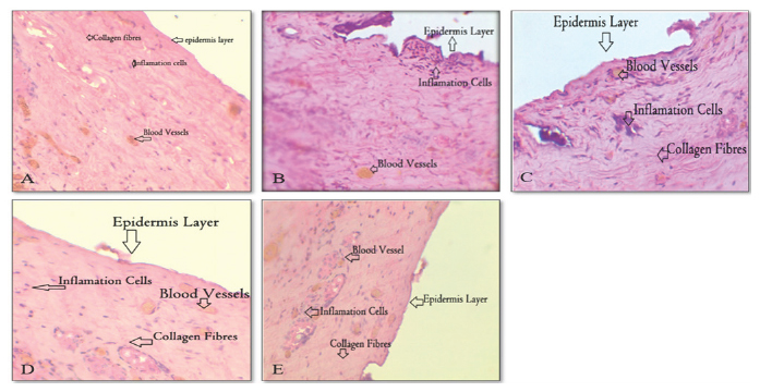
A: Standard Groups (STD)s (silver sulfadiazine cream) showing complete develop epithelial layer, less number of inflammation cells, more collagen fibers and fully formed blood vessels, B: Group 2:- Control Group (saline or vehicle only) Shown broken epithelial layer, more number of inflammation cells, less collagen fibers and dully formed blood vessel, C: Group 3:- Test 1 (5% Extract of MC) showing not properly developed epithelial layer, slightly less number of inflammation cells, slightly less collagen fibers compared to Group 1 and blood vessel s are lesser compared to Group1, D: Group 4:- Test 4 (Scaffold) showed properly developed epithelial layer then Group 3, more number of inflammation cells, collagen fibers compared to Group 3 and blood vessels also more compared to Group 3, E: Group 5:- Test 3 (5% Saponin Extract of MC) showing fully formed epidermis layer.
In comparison to rats with untreated hot wax burn wounds, the ninth day after administering the scaffold to the wound produced significant (p<0.01) wound constriction. Application of 5% Saponin of MC to the hot wax burn wound produced significant (p<0.001) wound contraction on the ninth day when compared to untreated hot wax burn wound rats.
Effect on Epithelization period (days)
Application of Silver Sulfadiazine Cream (1% w/w) to the hot wax burn wound significantly (p<0.001) reduced the epithelization period when compared to untreated hot wax burn wound rats. Whereas in Group 3, the hot wax burn wound significantly (p<0.01) reduced the epithelization period when compared to untreated hot wax burn wound rats. The epithelization period was not significantly (p<0.001) reduced on the application of the scaffold to the hot wax burn wound when compared to untreated hot wax burn wound rats. 5% Saponin of MC application significantly (p<0.001) reduced the epithelization period when compared to untreated hot wax burn wound rats (Table 1).
Total Protein
Silver Sulfadiazine Cream (1% w/w), methanolic extract MC (5% w/w/day), Scaffold, and 5% Saponin application to the hot wax burn wound produced a significant increase in total protein when compared to untreated hot wax burn wound rat (Figure 1 A and Table 2).
Hydroxyproline Content
Application of Silver Sulfadiazine Cream (1% w/w), scaffold, and 5% Saponin to the hot wax burn wound produced a considerable rise in the hydroxyproline level when compared to the untreated control. Whereas methanolic extract Momordica cymbalaira Fenzl. (5% w/w/day) did not produce a significant change in Hydroxyproline content (Figure 1B).
Antioxidant activity
Application of Silver Sulfadiazine Cream (1% w/w), methanolic extract MC. (5% w/w/day), Scaffold and 5% Saponin of MC to the hot wax burn wound produced a significant increase in Glutathione-s-transferase (Figure 2A), Catalase (CAT), (Figure 2B), Reduced Glutathione (GSH), (Figure 2C), activity in the granulation tissue and Superoxide Dismutase (SOD) (Figure 2D) when compared to untreated hot wax burn wound rats (Table 3).
| Groups | Wound Contraction (%) on the 9th Day of Topical Application | Epithelization Period (14 days) | ||
|---|---|---|---|---|
| 1stDay (Inch) | 9th Day (Inch) | Wound Contraction (9th days) (%) | ||
| Group 1 | 0.8 | 0.7 | 13.51 ± 1.95 | 18.00 ± 0.36 |
| Group 2 | 0.8 | 0.4 | 48.56 ± 1.04*** | 12.50±0.22*** |
| Group 3 | 0.8 | 0.6 | 24.58 ± 1.37** | 16.17 ± 0.40** |
| Group 4 | 0.8 | 0.6 | 24.00 ± 2.53** | 14.67±0.33*** |
| Group 5 | 0.8 | 0.5 | 33.25 ± 2.05*** | 14.17±0.31*** |
Effect of Methanolic Extract Momordica cymbalair Fenzl. (5% w/w/14 days), Saponin of MC (5% w/w/14 days), Scaffold, and Silver Sulfadiazine Cream (1% w/w/14 days) on Burn Wound on Topical Application
| Groups | Total protein (g/dl) | Hydroxyproline (mcg) |
|---|---|---|
| Group 1 | 0.8483 ± 0.12 | 2.700 ± 0.19 |
| Group 2 | 3.103 ± 0.10*** | 4.350 ± 0.74** |
| Group 3 | 1.235± 0.12*** | 3.683 ± 0.18 |
| Group 4 | 1.722 ± 0.17*** | 4.183 ± 0.2* |
| Group 5 | 2.242 ± 0.10*** | 4.783 ± 0.25*** |
Effect of Methanolic Extract Momordica cymbalaira Fenzl. (5% w/w/14 days), Saponin of MC (5% w/w/14 days), Scaffold, and Silver Sulfadiazine Cream (1% w/w/14 days) on Burn Wound on Topical Application
| Groups | Different Antioxidant Parameter | ||||
|---|---|---|---|---|---|
| Catalase (CAT) | Super oxide Dismutase(SOD) | Glutathione-S-Transferase (G-S-T) | Reduced Glutathione (GSH) | Lipid Peroxidase (MDA) | |
| Group 1 | 7.823± 0.2 | 1.302 ± 0.2 | 2.080 ± 0.3 | 19.38 ± 1.8 | 312.4 ±3.9 |
| Group 2 | 11.70±0.39*** | 15.36±0.55*** | 11.46±0.47*** | 82.71± 1.3*** | 114.5± 5.1*** |
| Group 3 | 9.223± 0.1*** | 2.692±0.2*** | 4.160±0.28*** | 32.92± 1.6*** | 261.3 ± 4.12*** |
| Group 4 | 9.197 ± 0.13*** | 3.743±0.19*** | 6.250± 0.21*** | 47.13± 2.30*** | 230.3 ± 2.9*** |
| Group 5 | 10.45± 0.2*** | 5.312± 0.4*** | 8.330± 0.3*** | 64.58± 1.8*** | 208.0 ± 4.9*** |
Effect of Methanolic Extract Momordica cymbalaira Fenzl. (5%w/w/14 days), Saponin of MC (5% w/w/14 days), Scaffold, and Silver Sulfadiazine Cream (1% w/w/14 days) on Burn Wound Induced Oxidative Stress in Granulation Tissue
Effect on Lipid Peroxidase (LP) (MDA)
Application of Silver Sulfadiazine Cream (1% w/w), methanolic extract Momordica cymbalaira Fenzl. (5% w/w/day), Scaffold, and 5% Saponin of MC to the hot wax burn wound produced a significant decrease in Lipid Peroxidase (MDA) activity in the granulation tissue when compared to untreated hot wax burn wound rats (Figure 2E).
Histological Structure of Granulation Tissue
5% Saponin Extract of MC and standard silver Sulphadiazine Cream showed a fully developed form of the epidermis layer, less inflammatory cells, more no. of collagen fibers, and blood vessels better against control groups. 5% Extract of MC and Scaffold also showed a full form of the epidermis layer, fewer inflammation cells, and more no. of collagen fibers and blood vessels compared to control but lesser than 5% Saponin Extract of MC and standard silver Sulphadiazine Cream. So, the Momordica cymbalaria
extracts increased the number of collagen and blood vessels in the wound site and also decrease inflammation cells (Figure 3).
DISCUSSION
Burn wound healing is a complex process that involves steps like coagulation, inflammation, matrix deposition, formation of new blood vessels, growth of fibrous tissue, epithelialization, contraction, and remodeling. The healing involves the intervention of the immune system that contributes to the remodeling process.38 In the present study, the scaffold has been employed. The scaffold can be seen as an alternate treatment method that either provides a source for the wound dressing or as a delivery system of the drug.
Wound contraction is a complicated procedure that necessitates the precise coordination of cells, extracellular matrix, and inflammatory mediators mainly cytokines.39 The fibroblast’s mobility promotes collagen and matrix production in the area of the wound. Present study results illustrated that in MC (Groups 3 and 4) and scaffold-treated (Group 5) groups, the % wound contraction was increased, which could be attributable to increased fibroblast activity in rejuvenated injured tissue.
In the current study, it was observed that MC saponins boosted levels of hydroxyproline in burn rats, increasing the collagen strength of the wound tissue that was regenerated.
The wound’s epithelialization period is the days required for complete epithelialization. In this stage, the keratinocyte drifts and then divides. Application of methanolic extract MC and 5% Saponin of MC significantly reduced the epithelialization period when compared to the control group. Whereas the scaffold did not produce any significant reduction in the epithelialization period.
After a burn injury, oxidative stress has a significant impact on the development of edema. It has been shown that there is a direct correlation between the degree of lipid peroxidation and postburn complications and that the ROS that causes lipid peroxidation plays a role in the mechanism of both local and systemic consequences in burns, including increased vascular permeability.40 MDA levels were observed to be much lower in the current investigation after Saponin of MC Fenzl treatment, indicating lessened oxidative injury. This discovery could be attributed to increased levels of antioxidants and their free radical scavenging property. All the groups except the first two showed a significant (p<0.001) increase in activity in the granulation tissue, Glutathione-S-Transferase (G-S-T), Catalase (CAT), Reduced Glutathione (GSH), Superoxide Dismutase (SOD).
Histopathological images illustrated that 5% Saponin Extract of MC showed complete epithelization a smaller number of inflammatory cells, and a greater number of collagen fibers and blood vessels, these results align with the results of the standard. 5% Extract of MC and scaffold showed similar results to a lesser extent when compared to the standard. Similar results were also observed with research conducted by Kumar and Kameshwarn et al. on extract and had a favorable impact on cellular proliferation, granular tissue development, and epithelization, according to earlier investigations on its therapy on dermal and epidermal regeneration.1,42
CONCLUSION
This study revealed that the administration of the methanolic extract, 5% saponin of Momordica cymbalaira Fenzl, and scaffold enhance burn wound healing in burn-induced animals. This was demonstrated by a substantial upsurge in the total protein, rate of wound contraction, hydroxyproline content, decreased Lipid Peroxidation (LP), and the epithelization period.
An increase in antioxidant activity was observed signifying the free radical scavenging property and their ability to destroy the reactive oxygen species.
Hence Momordica Cymbalaira Fenzl possesses burn wound healing activity by using the hot wax model in rats.
Cite this article
Nagashree KS, Solanki V, Koneri R, Nair SP, Chaithra SR. Evaluation of the Potential of
ACKNOWLEDGEMENT
We would like to thank JSS College of Pharmacy, Mysuru for their support throughout the study.
ABBREVIATIONS
| MC: | |
|---|---|
| SD: | Sprague-Dawley |
| LP: | Lipid Peroxidation |
| MDA: | Malondialdehyde |
| SOD: | Superoxide dismutase |
| CAT: | Catalase |
| GST: | Glutathione-s-transferase |
| GSH: | Reduced glutathione |
References
- Li Z, Maitz P. Cell therapy for severe burn wound healing. Burns Trauma. 2018;6:13 [PubMed] | [CrossRef] | [Google Scholar]

- Wang Y, Beekman J, Hew J, Jackson S, Issler-Fisher AC, Parungao R, et al. Burn injury: challenges and advances in burn wound healing, infection, pain and scarring. Adv Drug Deliv Rev. 2018;123:3-17. [PubMed] | [CrossRef] | [Google Scholar]

- Burns [internet]. [[cited Oct 27 2022]]. Available from: https://www.who.int/news-room/fact-sheets/detail/burns

- Tiwari VK. Burn wound: how it differs from other wounds?. Indian J Plast Surg. 2012;2012(2):364-73. [PubMed] | [CrossRef] | [Google Scholar]

- Werner S, Krieg T, Smola H. Keratinocyte-fibroblast interactions in wound healing. J Invest Dermatol. 2007;2007(5):998-1008. [PubMed] | [CrossRef] | [Google Scholar]

- Widgerow AD. Cellular/extracellular matrix cross-talk in scar evolution and control. Wound Repair Regen. 2011;2011(2):117-33. [PubMed] | [CrossRef] | [Google Scholar]

- Anand U, Jacobo-Herrera N, Altemimi A, Lakhssassi N. metabolites A Comprehensive Review on Medicinal Plants as Antimicrobial Therapeutics: potential Avenues of Biocompatible Drug Discovery. Available from: http://www.mdpi.com/journal/metabolites
[PubMed] | [CrossRef] | [Google Scholar]
- Aftab T, Hakeem KR. Medicinal and aromatic plants: expanding their horizons through omics. Secondary metabolites: an introduction to natural products chemistry. 2020 [PubMed] | [CrossRef] | [Google Scholar]

- Thakur R, Jain N, Pathak R, Sandhu SS. Practices in wound healing studies of plants. Evid Based Complement Alternat Med. 2011;2011:438056 [PubMed] | [CrossRef] | [Google Scholar]

- De A, Barbosa P. Pharmacologically active saponins from the genus Albizia (Fabaceae). Int J Pharm Pharm Sci. 2014:32-6. [PubMed] | [CrossRef] | [Google Scholar]

- Kimura Y, Sumiyoshi M, Samukawa Kichi, Satake N, Sakanaka M. [Facilitating action of asiaticoside at low doses on burn wound repair and its mechanism]. Eur J Pharmacol. 2008;584(2-3):415-23. [PubMed] | [CrossRef] | [Google Scholar]

- Liu M, Dai Y, Li Y, Luo Y, Huang F, Gong Z, et al. Madecassoside isolated from herbs facilitates burn wound healing in mice. Planta Med. 2008;2008(8):809-15. [PubMed] | [CrossRef] | [Google Scholar]

- Sharath R, Harish BG, Krishna V, Sathyanarayana BN, Swamy HM. Wound healing and protease inhibition activity of Bacoside-A, isolated from wettest. Phytother Res. 2010;2010(8):1217-22. [PubMed] | [CrossRef] | [Google Scholar]

- Song J, Dai Y, Bian D, Zhang H, Xu X, Xia Y, et al. Madecassoside induces apoptosis of keloid fibroblasts via a mitochondrial-dependent pathway. Drug Dev Res. 2011;2011(4):315-22. [CrossRef] | [Google Scholar]

- Rao BK, Kesavulu MM, Giri R, Appa Rao C. Antidiabetic and hypolipidemic effects of Hook. fruit powder in alloxan-diabetic rats. J Ethnopharmacol. 1999;1999(1):103-9. [PubMed] | [CrossRef] | [Google Scholar]

- Vrushabendra Swamy B, Jayaveera K, Reddy K, Swamy TB, Bharathi T. Anti-diarrhoeal activity of fruit extract of Hook F. Internet J Nutr Wellness. 2007;5(2) [PubMed] | [CrossRef] | [Google Scholar]

- Bhuvaneswari G, Thirugnanasampandan R, Ramnath MG. Evaluation of antioxidant, cytotoxic and apoptotic activities of methanolic extract of hook f unripe fruit. Kongunadu. 2020;2020(2):26-9. [CrossRef] | [Google Scholar]

- Chaitanya G, Pavani C, Suvarchala V, Bai DS, Spoorthi RD. Array. J Pharmacogn Phytochem. 2021;2021(4):146-52. [CrossRef] | [Google Scholar]

- Koneri R, Balaraman R, Saraswati CD. Antiovulatory and abortifacient potential of the ethanolic extract of roots of in rats. Indian J Pharmacol. 2006;2006(2):111 [CrossRef] | [Google Scholar]

- Pramod K, Deval RG, Lakshmayya RS, Ramachandra SS. Antioxidant and hepatoprotective activity of tubers of Momordica tuberosa Cogn. against CCl4 induced liver injury in rats undefined. Indian J Exp Biol. 2008;2008(7):510-3. [PubMed] | [Google Scholar]

- Echeverria Molina MI, Malollari KG, Komvopoulos K. Design challenges in polymeric scaffolds for tissue engineering. Front Bioeng Biotechnol. 2021;9:617141 [PubMed] | [CrossRef] | [Google Scholar]

- Negut I, Dorcioman G, Grumezescu V. Scaffolds for wound healing applications. Polymers. 2020;12(9):2010 [internet] [PubMed] | [CrossRef] | [Google Scholar]

- Alabdul Magid A, Voutquenne-Nazabadioko L, Renimel I, Harakat D, Moretti C, Lavaud C, et al. Triterpenoid saponins from the stem bark of . Phytochemistry. 2006;2006(19):2096-102. [PubMed] | [CrossRef] | [Google Scholar]

- Sánchez-Medina A, Stevenson PC, Habtemariam S, Peña-Rodríguez LM, Corcoran O, Mallet AI, et al. Triterpenoid saponins from a cytotoxic root extract of subsp. gaumeri. Phytochemistry. 2009;2009(6):765-72. [PubMed] | [CrossRef] | [Google Scholar]

- Larhsini M, Marston A, Hostettmann K. Triterpenoid saponins from the roots of . Fitoterapia. 2003;2003(3):237-41. [PubMed] | [CrossRef] | [Google Scholar]

- Lu J, Xu B, Gao S, Fan L, Zhang H, Liu R, et al. Structure elucidation of two triterpenoid saponins from rhizome of Regel. Fitoterapia. 2009;2009(6):345-8. [PubMed] | [CrossRef] | [Google Scholar]

- Nayak BS, Raju SS, Eversley M, Ramsubhag A. Evaluation of wound healing activity of L. -a preclinical study. Phytother Res. 2009;2009(2):241-5. [PubMed] | [CrossRef] | [Google Scholar]

- Sawant SE, Tajane MD. Formulation and evaluation of herbal ointment containing Neem and Turmeric extract. J Sci Innov Res. 2016;2016(4):149-51. [CrossRef] | [Google Scholar]

- Srivastava P, Durgaprasad S. Burn wound healing property of : an appraisal. Indian J Pharmacol. 2008;2008(4):144-6. [PubMed] | [CrossRef] | [Google Scholar]

- Chithra P, Sajithlal GB, Chandrakasan G. Influence of aloe vera on the healing of dermal wounds in diabetic rats. J Ethnopharmacol. 1998;1998(3):195-201. [PubMed] | [CrossRef] | [Google Scholar]

- Datta HS, Mitra SK, Patwardhan B. Wound healing activity of topical application forms based on Ayurveda. Evid Based Complement Alternat Med. 2011;2011:134378 [PubMed] | [CrossRef] | [Google Scholar]

- Durmus M, Karaaslan E, Ozturk E, Gulec M, Iraz M, Edali N, et al. The effects of single-dose dexamethasone on wound healing in rats. Anesth Analg. 2003;2003(5):1377-80. [PubMed] | [CrossRef] | [Google Scholar]

- Sakat SS, Juvekar AR, Gambhire MN. Array. [PubMed] | [CrossRef] | [Google Scholar]

- Elstner EF, Heupel A. Inhibition of nitrite formation from hydroxylammoniumchloride: a simple assay for superoxide dismutase. Anal Biochem. 1976;1976(2):616-20. [PubMed] | [CrossRef] | [Google Scholar]

- Ramanathan M, Sivakumar S, Anandvijayakumar PR, Saravanababu C, Pandian PR. Neuroprotective evaluation of standardized extract of in monosodium glutamate treated rats. Indian J Exp Biol. 2007;2007(5):425-31. [PubMed] | [Google Scholar]

- . Glutathione S-transferase activity of erythrocyte genotypes HbAA, HbAS and HbSS in male volunteers administered with Fansidar and quinine. Adv J Microbiol Res. 2009;2009(2):1-5. [PubMed] | [Google Scholar]

- Verma S, Samarth R, Panwar M. Evaluation of radioprotective effects of Spirulina in Swiss albino mice. 2006 [PubMed] | [Google Scholar]

- Oryan A, Alemzadeh E, Moshiri A. Burn wound healing: present concepts, treatment strategies and future directions. J Wound Care. 2017;2017(1):5-19. [internet] [PubMed] | [CrossRef] | [Google Scholar]

- Gupta A, Upadhyay NK, Sawhney RC, Kumar R. A poly-herbal formulation accelerates normal and impaired diabetic wound healing. Wound Repair Regen. 2008;2008(6):784-90. [PubMed] | [CrossRef] | [Google Scholar]

- Gupta VK. Assessment of antioxidant activity of in healing of thermal burn wound with and without supportive treatment of silver sulfadiazine in rabbits. Int J Basic Clin Pharmacol. 2019;2019(7):1594 [CrossRef] | [Google Scholar]

- Senthil Kumar M, Sripriya R, Raghavan H, Sehgal PK. Wound Healing Potential of on Infected albino Rat Model. J Surg Res. 2006;2006(2):283-9. [PubMed] | [CrossRef] | [Google Scholar]

- Kameshwara S, Senthilkum R, Thenmozhi S, Dhanalaksh M. Wound Healing Potential of ethanolic Extract of Flowers in Rats. Pharmacologia. 2014;2014(6):215-21. [CrossRef] | [Google Scholar]


